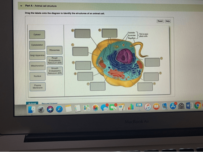
Drag the labels onto the diagram to identify cytoskeletal structures, an interactive learning activity, takes center stage, inviting learners into a captivating realm of cellular biology. This engaging experience empowers individuals to explore the intricate world of cytoskeletal structures, gaining a deeper understanding of their functions and significance within the cell.
As we delve into this activity, we will uncover the diverse types of cytoskeletal structures, including microtubules, microfilaments, and intermediate filaments. Through interactive exploration, learners will gain hands-on experience in identifying these structures and comprehending their roles in maintaining cell shape, facilitating movement, and organizing cellular processes.
Cytoskeletal Structures: Drag The Labels Onto The Diagram To Identify Cytoskeletal Structures

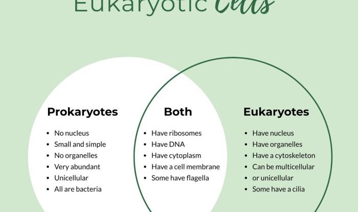
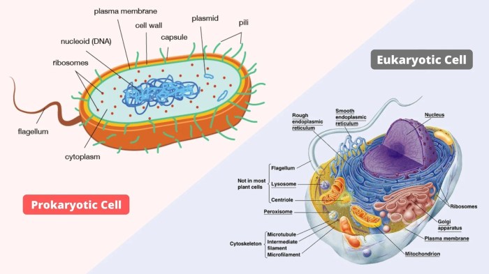
The cytoskeleton is a dynamic network of protein filaments and tubules that provides structural support and organization to eukaryotic cells. It plays a crucial role in various cellular processes, including cell shape maintenance, cell division, intracellular transport, and cell signaling.
There are three main types of cytoskeletal structures: microtubules, microfilaments, and intermediate filaments.
Microtubules
Microtubules are long, hollow, cylindrical structures composed of tubulin proteins. They are the thickest of the cytoskeletal structures, with a diameter of about 25 nm. Microtubules are responsible for maintaining cell shape, providing structural support, and facilitating intracellular transport. They also play a critical role in cell division, forming the mitotic spindle that separates chromosomes during cell division.
- Examples:Spindle fibers, cilia, and flagella
- Function:Cell shape maintenance, intracellular transport, cell division
Microfilaments
Microfilaments are thin, solid, filamentous structures composed of actin proteins. They are the smallest of the cytoskeletal structures, with a diameter of about 7 nm. Microfilaments are involved in cell shape changes, cell movement, and muscle contraction. They also play a role in cell division, forming the contractile ring that pinches the cell in two during cytokinesis.
- Examples:Actin filaments, stress fibers
- Function:Cell shape changes, cell movement, muscle contraction, cell division
Intermediate Filaments, Drag the labels onto the diagram to identify cytoskeletal structures
Intermediate filaments are intermediate in size between microtubules and microfilaments, with a diameter of about 10 nm. They are composed of various proteins, including keratin, vimentin, and desmin. Intermediate filaments are responsible for providing mechanical strength and stability to cells.
They also play a role in cell shape maintenance and cell-cell adhesion.
- Examples:Keratin filaments, vimentin filaments
- Function:Cell shape maintenance, cell-cell adhesion, mechanical strength
FAQ Compilation
What are the different types of cytoskeletal structures?
There are three main types of cytoskeletal structures: microtubules, microfilaments, and intermediate filaments.
What are the functions of cytoskeletal structures?
Cytoskeletal structures play crucial roles in maintaining cell shape, facilitating movement, and organizing cellular processes.
How does the drag-and-drop activity help in learning about cytoskeletal structures?
The drag-and-drop activity provides an interactive and engaging way to identify and learn about different types of cytoskeletal structures.