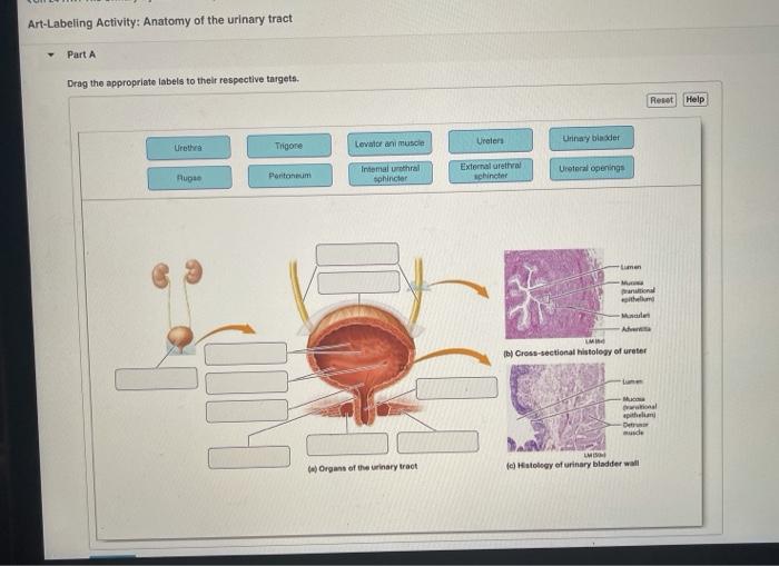
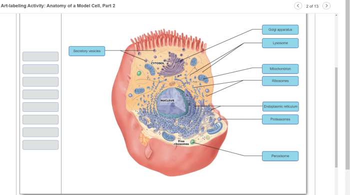
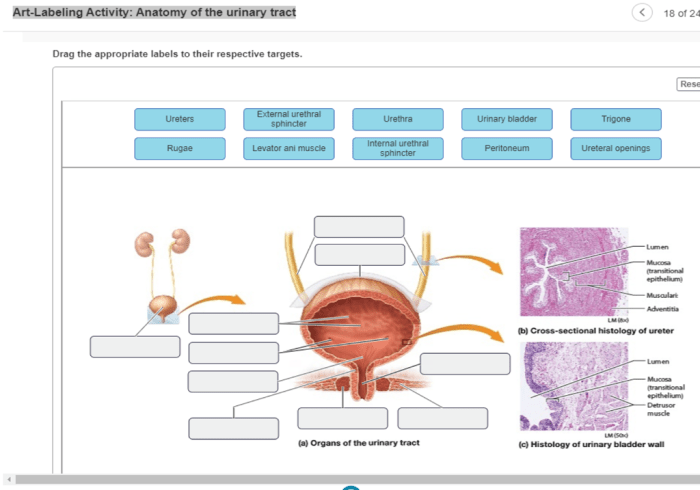
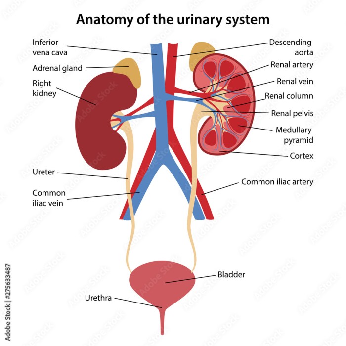
Unveiling the Art-Labeling Activity: Anatomy of the Urinary Tract, an innovative pedagogical approach that harnesses the power of visual representation to revolutionize the understanding of this complex anatomical system. This engaging activity not only enhances cognitive abilities but also fosters a deeper comprehension of anatomical structures and their intricate relationships.
Through a step-by-step guide, we delve into the art-labeling process, providing resources for obtaining anatomical illustrations and exploring various labeling techniques. By actively engaging with visual representations, students embark on a journey of discovery, reinforcing their understanding of the urinary tract’s intricate network.
1. Introduction: Art-labeling Activity: Anatomy Of The Urinary Tract
Accurate labeling is crucial in medical education and research. Understanding the anatomy of the urinary tract is essential for medical professionals to diagnose and treat urinary system disorders.
2. Art-Labeling Activity
Anatomy of the Urinary Tract
Step-by-Step Guide:
- Obtain anatomical illustrations of the urinary tract (e.g., from textbooks, online resources).
- Create or gather physical or online labels for the anatomical structures.
- Instruct students to match the labels to the correct anatomical structures on the illustrations.
- Provide feedback and guidance as needed.
3. Benefits of Art-Labeling Activities

Cognitive Benefits:
- Enhanced memory retention
- Improved spatial reasoning
- Increased understanding of anatomical relationships
Educational Applications:
- Successful use in medical education to improve comprehension of anatomical structures
- Reinforcement of textbook learning
4. Assessment and Evaluation

Assessment Methods:
- Accuracy and completeness of labeled structures
- Written or oral presentations explaining the labeled structures
Evaluation:
- Use assessment results to improve future art-labeling activities
- Provide specific feedback to students on their labeling accuracy
5. Incorporation into Curriculum

Integration into Medical Education:
- Incorporation into anatomy, physiology, and histology courses
- Use as a supplement to traditional teaching methods
Potential Benefits:
- Improved understanding of urinary tract anatomy
- Enhanced retention of anatomical information
- Increased engagement and motivation
6. Future Directions

Research and Development:
- Exploration of technology integration (e.g., virtual reality, augmented reality)
- Assessment of art-labeling activities in other anatomical systems
Potential Applications:
- Development of interactive anatomical learning tools
- Assessment of student understanding in various medical disciplines
Answers to Common Questions
How does art-labeling enhance anatomical understanding?
Art-labeling fosters a deeper comprehension of anatomical structures by engaging multiple senses and promoting active recall. The process of labeling illustrations reinforces knowledge, improves spatial reasoning, and cultivates a holistic understanding of the body’s intricate systems.
What are the benefits of incorporating art-labeling activities into medical curricula?
Integrating art-labeling activities into medical education offers numerous advantages. These activities enhance memory retention, develop critical thinking skills, and promote collaboration among students. Moreover, they provide a unique and engaging way to explore complex anatomical concepts, fostering a deeper understanding of the human body.